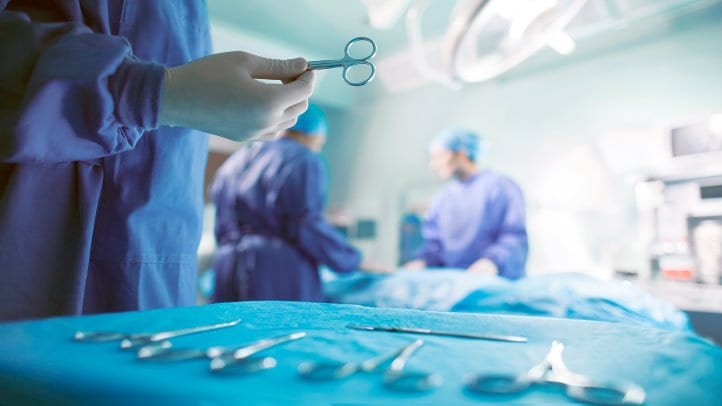

Ever since its inception in the mid-1980s, image-guided surgery (IGS) has revolutionized the face of medicine in several ways. It has tremendously changed the way surgeons execute difficult surgical procedures including the operation time, the precision of incisions, and the management of risk to healthy tissues. Through IGS, surgeons can plan how to make accurate procedures targeting abnormal tissue while avoiding surrounding critical structures.
According to a 2000 study, medical researchers had already anticipated that a huge portion of future neurosurgery would employ computer-based technologies. Well, it is now a reality, thanks to image-guided surgery. If you are considering surgery in the near future, you might benefit from learning how such computerized technologies have made surgery a safe and easy practice.
IGS was initially developed for neurosurgery but other applications such as endoscopic sinus surgery (ESS) are fast employing it for its vast benefits.
Here, we have outlined 5 leading applications of image-guided surgical systems, but first, let’s define IGS.
What is image-guided surgery?
The term image-guided surgery (IGS) refers to a surgical procedure that makes use of tracked surgical instruments with intraoperative images or preoperative images to navigate the procedure. With this technology, a surgeon links the operative field to the preoperative imaging data reflecting the exact location of a surgical instrument to the adjacent anatomic structures.
By registering anatomy models with the actual position of a patient in the operation table, one can give the surgeon augmented reality visualizations displaying the hidden tissue in exact alignment with a real-time view of the patient. Tracking the surgical instrument related to both the patient and the registered model provides real-time feedback about its position as well as its relationship to surrounding tissue.
The computer is undoubtedly the driving force behind the success of image-guided surgery. It serves many purposes in biomedical engineering including;
- Display and manipulation of medical images
- Control of imaging equipment
- Reconstruction of images from raw data
- Tracking instruments
- Modeling tissue
- Control of robots
- Evaluation of surgical performance
- Facilitation of human-machine interface
Benefits of Image-Guided Surgery
Image-guided surgery enhances precision enabling surgeons to create detailed plans of surgeries including appropriate incision spots, the correct path to the targeted region and sensitive structures to be avoided. It provides an opportunity for surgeons to view the human body in real-time 3D. With IGS, certain abnormalities like brain tumors can be clearly seen and distinguished from nearby tissue.
Image guided surgical systems also aids in shortening operating times and reducing the size of the incision. Both of these benefits enhance patient outcomes and fasten their recoveries. The technology also provides a solution for patients whose bodies may not stand invasive surgical procedures and those whose conditions would have otherwise been deemed intolerable.
Applications of Image-Guided Surgery
Orthopedics
One of the primary applications of IGS is in orthopedic surgeries specifically knee and hip replacements. This technique has been well adapted in Western Europe, especially Germany which happens to be the major market for image-guided orthopedic surgeries. There’s still a long way to go but surgeons are willing to perform computer-aided orthopedic surgeries to reduce errors and an array of complications.
Ear, Nose, and Throat (ENT)
ENT surgeries involve navigation of small anatomical cavities and structures. Add that to the limited visualization and you will understand why image-guided surgical systems come in handy in ENT. Some of the ENT procedures that make use of image-guided technology include cyst or polyp removal, sinus surgery, ENT surgery, reconstructive, skullbase surgery, and optic decompression. Thanks to the provision of 3D images of patients’ inner anatomy, Image guided surgical systems enhance better visualization than conventional 3D endoscopic procedures. Their less-invasive and precise characteristics improve outcomes, shorten recovery times and increase patient benefits through reduced risk. As awareness of the advantages of IGS in ENT continues to spread across the globe, more hospitals and clinicians appreciate its value and embrace the technology.
Neurology and neurosurgery
As mentioned earlier, image-guided surgical systems were introduced for use in neurosurgery. Over the years, many neurological procedures have heavily relied on image guidance involving preoperative images as well as intraoperative navigation. One of the branches of neurology where IGS has been primarily used is cranial applications. Currently, this technology is the standard for most of the surgical cranial operations in Western Europe. With the high risk associated with brain surgeries, IGS has provided surgeons with the ability to reduce the risks like damaging healthy tissues and instead increase surgical accuracy. This has allowed many of them to gain acceptance and credibility in the market. Spinal applications, too, employ IGS but these are yet to pick up the pace as the market keeps opening up.
Sentinel Lymph Node Mapping
Part of the emerging applications of image-guided surgeries includes sentinel lymph node mapping. The sentinel lymph node refers to the first lymph node that receives lymphatic fluid draining from the tumor. It is also the location where tumor cells begin to metastasize. Presently, lymphatic imaging uses such technologies as nuclear imaging, CT, dye-injection, and MRI. All these come with a host of limitations surrounding resolution, sensitivity, practical use, or exposure to radioactivity. IGS is a safe and simple technique that allows for increased spatial and temporal resolution devoid of ionizing radiation. When combined with infrared light, the outcome is a clear distinction of the sentinel lymph nodes from the surgical field.
Optimal IGS
The development of intra-operative optical camera imaging systems for the detection of tumors in mice during surgery is currently underway. Additionally, several endoscopic systems are also being developed for both surgical and diagnostic applications. One of the recent studies in the application of IGS for neurosurgery is the use of 5-aminolevulinic acid that aid in the detection of malignant gliomas using optical imaging techniques.
Image-guided surgery may not be a worldwide medical phenomenon now but it is certainly influencing the way medical procedures are performed. With computers offering valuable help, future surgical procedures are poised to be less invasive, shorter, more successful, and pose little or no risk to patients.